When one makes the decision to have Laser-Assisted In Situ Keratomileusis (LASIK), they look forward to having fantastic eyesight. They have visions of seeing the leaves on the trees, throwing away their glasses and contact lenses, and waking up with the ability to see their alarm clock.
Sometimes the results of LASIK start off this way, but then the vision starts to slip away. The blur, double vision, glare, halos, shadows, and eyestrain return. What happened to the crisp vision that was a reality for just a short time? Perhaps it’s corneal ectasia. Among the various LASIK complications, corneal ectasia can be one of the most devastating.
During LASIK, a protective flap is created in the cornea. This flap is then lifted and the laser treatment delivered. The flap is then replaced to help the cornea heal.
Prior to undergoing the procedure, a patient’s candidacy must be evaluated. Part of this assessment involves the measurement of corneal thickness or pachymetry. Pachymetry is an essential screening measurement, as it helps to determine the safety of a laser procedure. The laser treatment, or ablation, removes corneal tissue; therefore, there must be enough there to begin with so that sufficient tissue is left post-operatively. If the cornea is too thin, removing tissue could leave the cornea weakened and prone to distortion.
Sufficient pachymetry readings are especially important when a patient has a high glasses prescription, as this means more tissue must be removed to fully correct their vision. Similarly, if a patient has had LASIK before, they could be at an increased risk of a weakened cornea with further treatment. Therefore, caution must be used with these patients.
Another part of screening for LASIK involves mapping the cornea with topography. There are certain key aspects of topography that can help to assess for LASIK safety as well.
If the cornea is too thin and/or too much tissue is removed, ectasia could result. Ectasia is a bulging of the cornea that results from the cornea being structurally compromised. When the cornea is too thin, it doesn’t have the strength to maintain its shape. Over time, the cornea forms a protrusion, usually inferiorly, called ectasia. This area of the cornea is steep in curvature relative to its surrounding tissue.
When the cornea with post-LASIK ectasia is imaged using a mapping system called topography, the ectatic area will be shaded in warm colors, indicating steepness. Post-LASIK ectasia is similar to a naturally occurring condition called keratoconus. In fact, if a subtle case of keratoconus is missed prior to LASIK, the risk of ectasia increases. Corneal ectasia generally develops months to a year or two after LASIK.
Because the corneal surface is so irregular in shape in ectasia, light is scattered when it hits the eye. Corneal ectasia symptoms include blur, double vision, glare, halos, eyestrain, and headaches. These symptoms are not easily treated with glasses or soft contact lenses. While gas-permeable contact lenses can sometimes be an option, comfort may be a limiting factor. Plus, many patients first underwent refractive surgery to get away from wearing contacts!

Thankfully, there are now surgical techniques for corneal ectasia repair. Only a handful of surgeons, such as Dr. Motwani, are trained in laser corneal reconstruction. Dr. Motwani is able to smooth out the surface of the cornea using laser ablation.
Dr. Motwani uses Wavelight Contoura corneal topography to guide this procedure. This technology is approved by the Food & Drug Administration (FDA) for the treatment of corneal irregularity. This powerful corneal topographic system allows him to target the correction of individual points on the cornea. This enables the restoration of an ideal corneal shape for sharp vision.
Corneal crosslinking (CXL) is a procedure that improves the strength of a weakened cornea. It involves first sloughing away the outermost corneal cells. Riboflavin eye drops are then placed in the eye. Finally, ultraviolet light is applied. This combination of riboflavin with ultraviolet light results in a strengthening of the collagen layers in the cornea. CXL is valuable for use after surgical repair of the cornea, as it serves to lock the result in place. The cornea is cured in its desired shape to avoid shifts post-surgically.
With corneal ectasia, the tissue is prone to changing shape even after it has been treated with laser corneal reconstruction. The use of corneal cross-linking is a powerful tool to lock the results in place. The combination of laser corneal reconstruction and corneal cross-linking allows for a lasting corneal restoration.
Wouldn’t it be nice to have the vision you were aiming for when you first had LASIK? You don’t have to suffer from corneal ectasia symptoms any longer. With laser corneal reconstruction and corneal cross-linking, Dr. Motwani can help you regain the eyesight you once had.
Cataract Surgery is the most performed surgical procedure in the United States, and has a phenomenal track record for improving…
Read More
The treatment of trauma with topographic-guided ablation depends on the level of scarring caused by the trauma, the position of…
Read More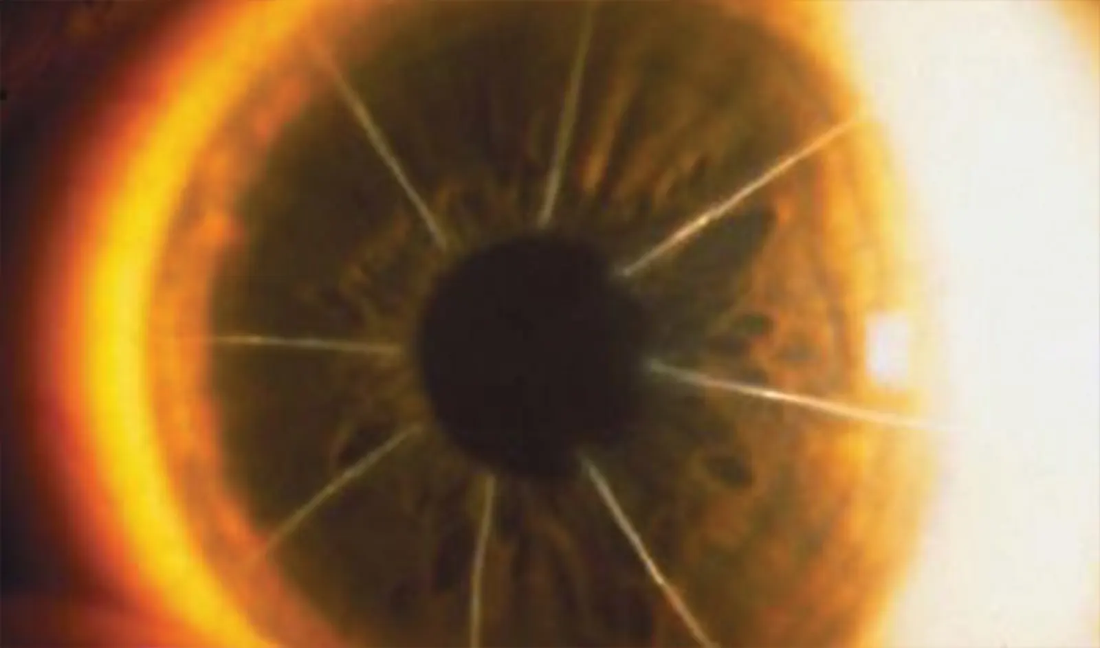
In the dynamic world of eye care, keratoconus treatment has become a focal point due to the condition’s impact on…
Read More