Radial Keratotomy was a procedure performed prior to laser vision correction, achieving the correction of myopia by diamond knife cut radial slices in the cornea to collapse the center of the cornea to achieve the refractive effect. These cuts are surgeon made by a template, and are 90% of the thickness of the cornea. It is very common for the cornea to end up with significant irregularity, causing a change in refraction as well as distortion of the vision.
Correction with topographic guided ablation corrects the irregularity, making for not only more stable vision, but can result in better quality vision with decreased distortion. Often the correction of the irregularity can correct a large portion of the refractive correction of the eye, and then fine-tuned with an enhancement for full final correction and better structure.
We have a long history of performing correction of radial keratotomy, dating back to 2001. We will treat these corrections in most cases utilizing LASIK, so downtime is minimal. Enhancements can also be performed utilizing the same LASIK flap via a technique pioneered by Dr. Motwani, and downtime is again minimal.
Topographic guided ablation (Contoura) of RK utilizing the San Diego Protocol created and published by Dr. Motwani has been very successful at treating even extremely severe irregularities of the cornea.
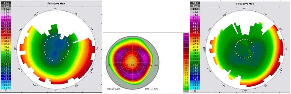
71 year old male; OD T-CAT PRK
Pre-op Manifest; OD: +1.50-1.00X123 BCVA 20/20
T-CAT Treatment; OD: +0.00-1.43X10⁷
3 Month Post Op; OD Refraction: plano and 20/20
OU vision: 20/20

71 year old male; OS T-CAT + WFO PRK
Pre-op Manifest; OS: +1.75 BCVA 20/20
T-CAT Treatment; OS: +0.00-0.61X128
WFO Treatment; OS: +1.00
3 Month Post Op; OS Refraction: plano -0.50X014 and 20/20
OU vision: 20/20

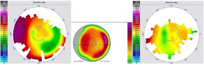
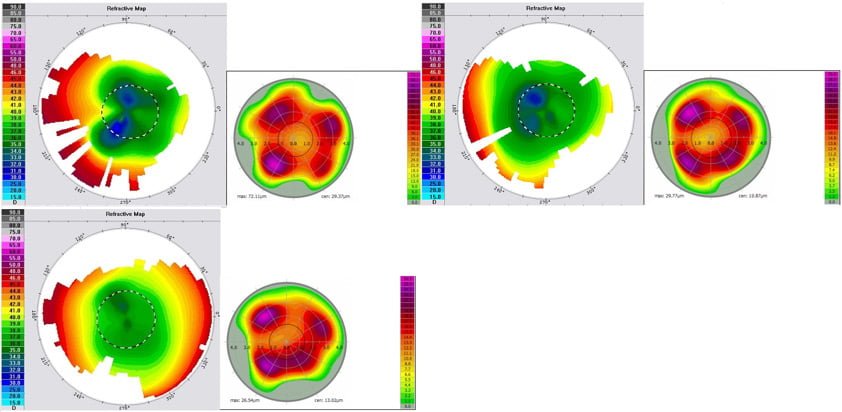
CASE 1
This is a 71 year old male who had Radial Keratotomy (RK ) performed in approximately 1994 in both eyes. He had LASIK performed in both eyes in 2004 with a successful correction. Patient returned in 2016 with a correction necessary again. Both eyes showed irregular astigmatism from the RK. Patient had irregular corneal astigmatism repair attempted with total refractive correction with one procedure in both eyes, utilizing the Contoura measured refraction, and both eyes achieved 20/20 vision. Interestingly, the patient at 6 months stated his vision was the best that he could ever remember it. The optics were subjectively better than with correction in the past with LASIK, RK, or glasses.
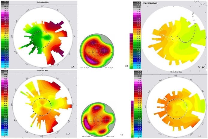
66 year old male, T-CAT LASIK OD
Pre-Op Manifest: OD: +3.75-1.75X075 20/20
T-CAT Corneal Equalization ONLY: OD: +0.00-0.60X094
ESX WFO OD at 3 months post-op: +3.00 D/S
3 Month Post Op; OD Refraction: +0.50 D/S and 20/20
OU vision: 20/20

66 year old male, T-CAT LASIK OS
Pre-Op Manifest: OS: +4.25-1.75X081 20/25
T-CAT Corneal Equalization ONLY: OS: +0.00-0.14X116
ESX WFO OS 3 months post-op: +2.75-0.75X090
3 Month Post Op; OS Refraction: plano and 20/30
OU vision: 20/20
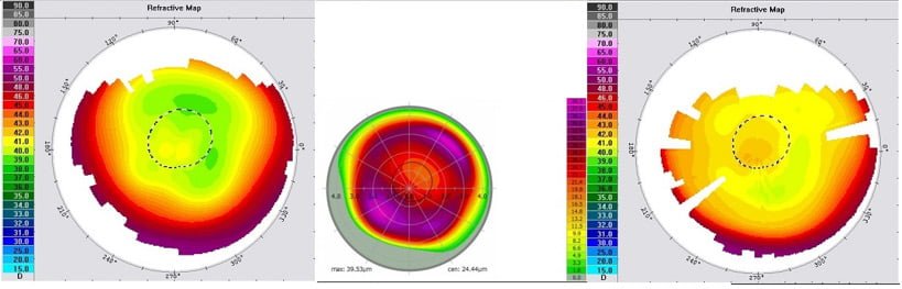
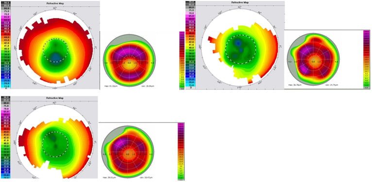
66 y.o. male with Radial Keratotomy (RK) performed approximately 20-25 years ago, patient believes somewhere around 1992. Pt. had a history of amblyopia OS from lazy eye, had strabismus surgery as a child. Patient had significant corneal irregularity causing visual difficulty in both eyes.
LASIK performed OU, with only correction of irregularity and Contoura Measured astigmatism. 3 months post-op, both eyes has final Wavefront Optimized correction resulting in 20/25 vision best corrected vision OU, with the left eye set up for reading via monovision.
67 year old male; OD T-Cat LASIK
Pre-Op Manifest; OD: +5.00-3.75X075 BCVA 20/25*
T-cat Treatment; OD: +0.00-3.00X078
ESX WFO OD; +2.00-2.25X110
3 month Post Op; OD Refraction: +0.50-0.75×095 and 20/25

67 year old male; OS T-Cat LASIK
Pre-Op Manifest; OS: +3.00-3.00X080 BCVA 20/25*
T-cat Treatment; OS: +0.00-2.00X087
ESX WFO OS; +1.00-0.75X040
3 month Post Op; OS Refraction: -0.75 D/S and 20/25 OU: 20/25
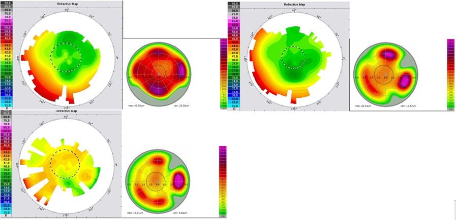
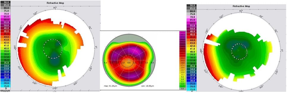
67 y.o. male suffering from frequent severe headaches. He had Radial Keratotomy (8- slit RK) in 1992. Pt. had severe irregularity with de-centration of central optical zone created by the RK.
Pt. had LASIK performed correcting the irregularity and Contoura Measured Astigmatism in both eyes. In the right eye, this eliminated 3.0D of hyperopia. Wavefront Optimized correction was performed at 3 months with resultant 20/25 vision.
In the left eye correcting the irregularity and the CMA eliminated 2.0D of hyperopia. After WFO enhancement at 3 months he had 20/25 vision.
Pt has also had almost complete elimination of his severe headaches.
80 year old male, OD T-CAT
Pre-op Manifest: OD: -3.25-1.00×090 BCVA 20/200
T-CAT Treatment: OD: -2.20-1.28X084
1 Month Post-OP: OD Refraction: plano and 20/40
80 y.o. male who had Radial Keratotomy (10-slit) RK in the 1970’s in the right eye. His best corrected vision was 20/200 with manifest refraction, and 20/70 with his RGP lens. Pt never had 20/20 vision in that eye-hx of amblyopia.
Right eye LASIK was performed with irregularity correction and refractive correction attempted at the same time. At 2 months post-op patient was 20/40 uncorrected with plano refraction.
49 year old male, T-CAT OD PRK
Pre-Op Manifest: OD: +2.25-0.75X095 BCVA 20/25
T-CAT Corneal Equalization Only: OD: +0.00-0.69X111
1 Month Post Op: OD Refraction: plano and 20/30 J3
49 year old male, T-CAT OS PRK
Pre-Op Manifest: OS: +3.00-0.50X030 BCVA 20/25
T-CAT Corneal Equalization Only: OS: +0.00-1.85X073
1 Month Post Op: OS Refraction: plano and 20/30 OU 20/20
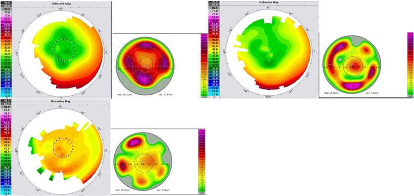
49 y.o. male with 16 slit Radial Keratotomy (RK) performed in 1986. Pt had PRK 8 years later at age 26, then PRK enhancement at 30. He had another LASEK performed at 36, PRK at 37, and PRK at 44.
Patient still had irregularity from the original RK. PRK was performed with corneal irregularity repair and only Contoura Measured Astigmatism correction in both eyes. Right eye post-op one month post-op was plano with 20/30 vision, and left eye was 20/30 with +1.00 vision. Equalizing the cornea eliminated 2.25D and 2.0D respectively by one month post-op.
69 year old male, OS T-CAT
Pre Op Manifest Rx: +6.75, -3.75 x 60 with BCVA of 20/30
Pre-Op Measured Astigmatism: -2.54 x 4
Treatment: 0 diopters sphere, 0 diopters cylinder (pure repair, no refractive error treated at all)
Post-Op 1 Day Initial Repair T-CAT Correction: 20/25 with refraction of +1.50
Pre-Enhancement Rx (3.5 months post-op initial correction): +1.25, -0.75 x 53
Measured Treatment: T-CAT– plano, – 2.59 x 17; WFO — +0.75
C4/12 compensation: +1.3 Diopters; Compensation for astigmatism increase, +0.92 Diopters; +2.22 – +1.25 = +0.97; clinical decision made to treat +0.75 and leave patient slightly hyper as patient wanted distance vision to be excellent.
Post Op Enhancement 1 month: plano and 20/20
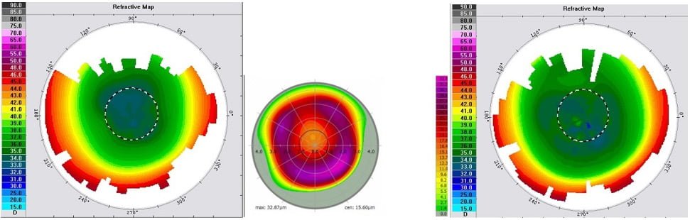
69 y.o. old male surgeon who had Radial Keratotomy (RK) in the left eye to attempt to treat hyperopia in the 1980’S. He ended up with severe irregularity that he felt had become career threatening.
We performed LASIK with only corneal irregularity repaired, which took 19 seconds of laser time with removal of 81 microns of tissue. This eliminated approximately 5.5D of hyperopia.
He had follow up Contoura treatment at 3 months that treated the Contoura Measured astigmatism and residual hyperopia resulting in 20/20 vision. This was a significant improvement over his best corrected 20/30 vision.
Cataract Surgery is the most performed surgical procedure in the United States, and has a phenomenal track record for improving…
Read More
The treatment of trauma with topographic-guided ablation depends on the level of scarring caused by the trauma, the position of…
Read More
In the dynamic world of eye care, keratoconus treatment has become a focal point due to the condition’s impact on…
Read More